The following extract is taken from the information provided by Small Animal Surgery Specialist Aidan McAlinden in the free webinar ‘Trauma – Spotting the Silent Killers’.
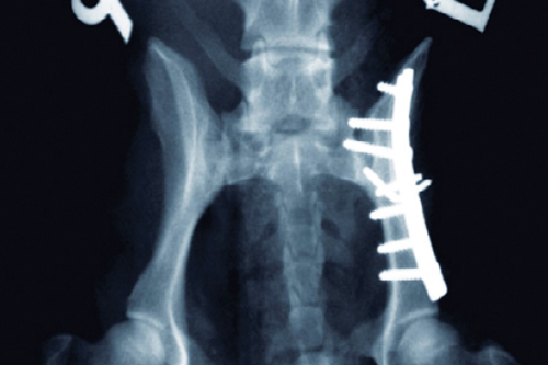
When investigating thoracic trauma patients, radiography is useful, but is not part of the emergency assessment. Animals may well decompensate due to the stress of trying to position them on an X-ray table, and so radiography should only be attempted as part of the diagnostic investigation after stabilisation of the patient. Pre-oxgenation and diagnostic thoracocentesis is urged in the first instance – then consider radiography later on.
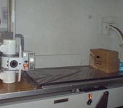
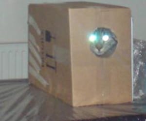
Cats are a special challenge. They are difficult to anaesthetise when they have significant respiratory stress. Even handling them to catheterise can be a challenge. The ‘Cat in a Box’ technique is really useful in these cases. Take a cardboard box and cut a small hole out of the front. Then put the cat in the box! Invariably, if they are reasonably alert, the cat will stand up to look out through the hole. Put a cassette at the side of the box, then drop the X-ray beam down to take a horizontal view. Obviously this is dependent on having lead lined walls and appropriate safety facilities for taking a horizontal beam radiograph. This can give us a really nice lateral view with minimal restraint of the cat and can be a real help in those difficult cases.
To get plenty more help with your emergency patients, you can watch the free webinar ‘Trauma – Spotting the Silent Killers’ by registering for free Silver Membership of Webinar Club and selecting ‘Free Webinars for Vets’ when you are logged in to the Members’ Area.
Find out more about the Emergency Surgery online Mini Series.